PRODUCTS SOLD ON PEPTIDESLABIRELAND.COM ARE FOR RESEARCH PURPOSES ONLY AND ARE NOT FOR HUMAN OR VETERINARY USE.
€196.00
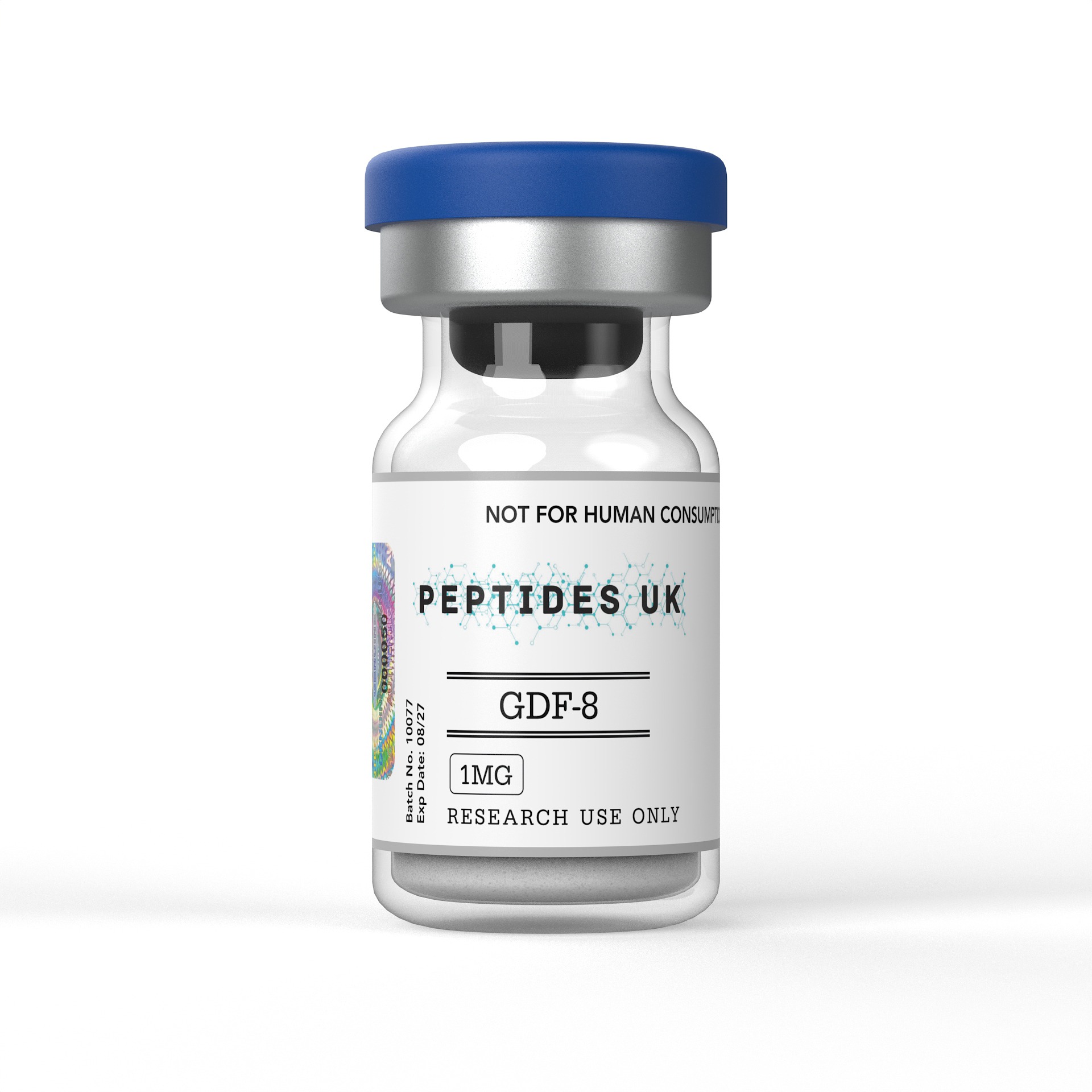
GDF-8 Ireland – Buy Online | In Stock & Ready to Ship
Buy GDF-8 in Ireland with fast shipping and guaranteed ≥99% purity — verified with COA and HPLC documentation. A trusted choice for peptides Ireland research teams rely on, with no customs delays or international wait times. Whether you’re searching for GDF-8 Ireland suppliers or looking to buy peptides Ireland-wide, we have you covered. Irish research teams can count on consistent stock, rapid fulfilment and full batch documentation every time.
For research use only. Not intended for human or veterinary use.


GDF-8 — Growth Differentiation Factor 8, more widely designated myostatin — is a synthetic recombinant TGF-beta superfamily member and one of the most biologically and therapeutically significant muscle regulatory research compounds available to laboratories in Ireland — a potent negative regulator of skeletal muscle mass whose signalling through ActRIIB/ALK4/ALK5 receptor complexes and downstream Smad2/3 transcriptional pathway suppresses myoblast proliferation, inhibits myogenic differentiation, promotes protein degradation, and maintains skeletal muscle mass within defined physiological limits, making it an indispensable research tool for studying myostatin receptor pharmacology and TGF-beta superfamily signal transduction, skeletal muscle atrophy and hypertrophy biology, myoblast proliferation and differentiation regulation, the ActRIIB-Smad2/3 signalling axis in muscle wasting disease models, satellite cell biology and muscle regeneration, the interaction between myostatin and follistatin in muscle mass regulation, metabolic consequences of muscle mass modulation including insulin sensitivity and energy expenditure, and the rapidly expanding research into myostatin pathway inhibition as a strategy for reversing muscle wasting in cachexia, sarcopenia, muscular dystrophy, and metabolic disease. Researchers and institutions across Ireland can source verified, research-grade GDF-8 directly from our Irish peptide supply, with domestic-speed dispatch and complete batch documentation.
✅ ≥99% Purity — HPLC & Mass Spectrometry Verified
✅ Batch-Specific Certificate of Analysis (CoA) Included
✅ Sterile Lyophilised Powder | GMP Manufactured
✅ Fast Dispatch to Ireland | Peptides Ireland Stock
GDF-8 — Growth Differentiation Factor 8 — is a member of the transforming growth factor beta (TGF-beta) superfamily of secreted signalling proteins, first identified and characterised by Alexandra McPherron and Se-Jin Lee at Johns Hopkins University in 1997 through positional cloning of the gene responsible for the extraordinary muscle hypertrophy observed in myostatin-null mice — animals with targeted disruption of the GDF8 gene that develop muscle masses two to three times greater than wild-type littermates through a combination of muscle fibre hyperplasia and hypertrophy. The GDF8 gene encodes a 375-amino acid prepropeptide that undergoes proteolytic processing to generate the mature GDF-8 homodimer — a disulfide-linked dimeric protein sharing the characteristic cystine knot structure of TGF-beta superfamily members — which circulates in plasma in both latent complexes with its propeptide and in active form capable of receptor engagement and downstream signalling.
The biological significance of GDF-8 as a research compound rests on its establishment as the primary endogenous negative regulator of skeletal muscle mass — a physiological brake on muscle growth whose genetic or pharmacological disruption produces dramatic and sustained muscle hypertrophy across multiple mammalian species including mice, cattle, sheep, dogs, and humans. Naturally occurring loss-of-function mutations in the GDF8 gene have been identified in Belgian Blue and Piedmontese cattle — breeds characterised by extreme muscularity or “double muscling” — and in rare human individuals with myostatin loss-of-function mutations who show extraordinary skeletal muscle development from birth. These naturally occurring myostatin-deficient phenotypes across multiple species established myostatin as a conserved and physiologically dominant regulator of muscle mass and identified the myostatin signalling pathway as a high-value target for research into muscle wasting diseases where pharmacological myostatin inhibition could restore or maintain muscle mass.
GDF-8 signals through a well-characterised receptor complex — binding with high affinity to the type II activin receptor ActRIIB on the surface of muscle cells, which then recruits and transphosphorylates the type I receptors ALK4 or ALK5, initiating downstream Smad2 and Smad3 phosphorylation, Smad2/3-Smad4 complex formation, nuclear translocation, and transcriptional regulation of target genes that suppress myoblast proliferation, inhibit the myogenic regulatory factors MyoD and myogenin, activate the E3 ubiquitin ligases MuRF1 and atrogin-1 to promote protein degradation, and collectively drive the muscle atrophy programme that myostatin physiologically maintains. Synthetic recombinant GDF-8 used in research reproduces the signalling biology of endogenous myostatin with equivalent receptor binding and downstream pathway activation — enabling mechanistic dissection of each step of the ActRIIB-Smad2/3 cascade and its consequences for muscle cell biology.
In controlled laboratory and pre-clinical settings, GDF-8 is studied across a range of myostatin receptor pharmacology, skeletal muscle atrophy biology, myoblast differentiation, satellite cell regulation, metabolic disease, and muscle wasting disease research applications:
Recombinant GDF-8 is the primary reference ligand for studying ActRIIB receptor pharmacology and downstream TGF-beta superfamily signal transduction in muscle biology — examining ActRIIB and ALK4/ALK5 receptor binding kinetics and affinity characterisation, type II to type I receptor recruitment and transphosphorylation, Smad2 and Smad3 C-terminal phosphorylation and activation, Smad2/3-Smad4 heteromeric complex assembly, nuclear translocation of activated Smad complexes, and transcriptional regulation of myostatin target genes including the myogenic regulatory factors, atrophy-associated ubiquitin ligases, and cell cycle regulators. Research has used GDF-8 to characterise structure-activity relationships within the TGF-beta superfamily relevant to ActRIIB engagement, to study how the type II-type I receptor co-complex formation and transphosphorylation cascade operates in muscle cell contexts, and to establish the complete signalling pathway from extracellular GDF-8 binding to nuclear transcriptional output. These receptor pharmacology studies have provided the molecular framework for understanding how a single TGF-beta superfamily ligand produces such pervasive effects on skeletal muscle mass regulation.
GDF-8 is extensively used in skeletal muscle atrophy research — where recombinant myostatin treatment of cultured myotubes, primary myoblasts, and satellite cells provides a controlled model for studying the molecular mechanisms of muscle protein degradation and atrophy programme activation. Research has used GDF-8 to characterise the complete atrophy gene programme activated downstream of Smad2/3 signalling — including transcriptional upregulation of the E3 ubiquitin ligases MuRF1 (muscle RING-finger protein 1) and atrogin-1/MAFbx that target myofibrillar proteins and myogenic regulators for proteasomal degradation, activation of the autophagy-lysosomal pathway that contributes to bulk protein and organelle degradation in atrophying muscle, suppression of the IGF-1/PI3K/Akt/mTOR anabolic signalling axis that normally maintains muscle protein synthesis, and inhibition of myogenic regulatory factor activity that suppresses the myogenic programme in mature muscle. These muscle atrophy mechanism studies have established the GDF-8-Smad2/3 axis as the primary driver of the transcriptional atrophy programme and GDF-8 as the essential research tool for activating and studying this programme in controlled experimental conditions.
GDF-8’s inhibitory effects on myoblast proliferation and myogenic differentiation represent a primary area of muscle cell biology research — with studies characterising how myostatin receptor activation suppresses the transition from proliferating myoblasts to post-mitotic differentiating myocytes that is essential for muscle fibre formation and regeneration. Research has used GDF-8 to examine the cell cycle regulatory mechanisms through which myostatin inhibits myoblast proliferation — documenting upregulation of the cyclin-dependent kinase inhibitors p21 and p27 and suppression of CDK2 activity that arrests myoblasts in G1 phase. Studies have characterised GDF-8’s inhibition of MyoD and myogenin transcriptional activity — the master myogenic regulatory factors whose sequential activation drives the commitment and differentiation programme converting myoblasts to myocytes — and the Smad2/3-dependent mechanisms through which myostatin receptor activation suppresses these myogenic regulators. These myoblast biology studies have established the dual mechanism through which GDF-8 limits muscle fibre formation — simultaneously suppressing proliferative expansion of the myoblast pool and blocking differentiation of those myoblasts that reach the differentiation commitment point.
Satellite cells — the resident adult muscle stem cell population residing beneath the basal lamina of mature muscle fibres — are the primary cellular mediators of skeletal muscle regeneration following injury, and myostatin is a critical regulator of satellite cell quiescence, activation, and self-renewal. Research has used GDF-8 to study how myostatin receptor activation maintains satellite cell quiescence in uninjured muscle, suppresses satellite cell activation and proliferative expansion following muscle injury, and modulates the balance between satellite cell self-renewal and commitment to differentiation during the regeneration process. Studies have characterised GDF-8’s effects on satellite cell behaviour in primary satellite cell cultures and in explanted muscle fibre models — examining how myostatin signalling through ActRIIB and Smad2/3 regulates the expression of quiescence markers, activation signals, and myogenic commitment factors in satellite cells. These satellite cell biology studies have contributed to understanding of how myostatin limits the regenerative capacity of adult skeletal muscle and how myostatin pathway inhibition could enhance muscle regeneration in injury and disease contexts.
GDF-8 is a central research tool in muscle wasting disease biology — where myostatin pathway activation contributes to the skeletal muscle atrophy characterising cachexia, sarcopenia, muscular dystrophy, spinal muscular atrophy, and amyotrophic lateral sclerosis. Research has used recombinant GDF-8 to establish in vitro atrophy models in muscle cell lines and primary myotubes that recapitulate the molecular features of disease-associated muscle wasting — allowing mechanistic dissection of atrophy pathway activation and screening of potential myostatin inhibitory strategies. Studies have examined how GDF-8-induced atrophy in cultured muscle cells models the pathological muscle wasting observed in cancer cachexia, ageing-associated sarcopenia, and Duchenne muscular dystrophy — characterising the shared molecular mechanisms including MuRF1/atrogin-1 upregulation, mTOR suppression, and myogenic regulatory factor inhibition that are common to disease-associated wasting and GDF-8-induced experimental atrophy. These disease model studies have established GDF-8 as the pharmacological tool for creating controlled muscle atrophy conditions in vitro that are mechanistically representative of pathological muscle wasting.
GDF-8’s role as the target of multiple endogenous and pharmacological inhibitors has generated a substantial research programme examining the biology of myostatin pathway inhibition — with follistatin, FLRG, GASP-1, and the myostatin propeptide each characterised as endogenous negative regulators of myostatin activity. Research has used recombinant GDF-8 as the activating ligand in competitive inhibition studies examining the binding affinities, inhibition kinetics, and functional potencies of these endogenous inhibitors — characterising the molecular basis for follistatin’s neutralisation of myostatin through direct high-affinity binding that prevents ActRIIB engagement. Studies have also used GDF-8 as the reference activating stimulus in screening assays for pharmacological myostatin inhibitors — including anti-myostatin antibodies, ActRIIB decoy receptors, and small molecule inhibitors of the Smad2/3 pathway — establishing GDF-8 as the essential positive control ligand for myostatin inhibitor characterisation research.
The metabolic consequences of myostatin signalling extend beyond skeletal muscle mass regulation to encompass insulin sensitivity, glucose homeostasis, and adipose tissue biology — reflecting the central role of skeletal muscle as the primary site of insulin-stimulated glucose disposal and the metabolic consequences of muscle mass changes on systemic energy metabolism. Research has used GDF-8 to study how myostatin receptor activation in muscle cells modulates insulin signalling — examining GDF-8’s inhibitory effects on the IGF-1R/IRS-1/PI3K/Akt/GLUT4 insulin signalling pathway in muscle cells, the consequences of myostatin-induced muscle mass reduction for systemic insulin sensitivity in metabolic disease models, and the relationship between myostatin pathway activation and the development of insulin resistance in obesity and type 2 diabetes. Studies have also examined myostatin’s effects on adipogenesis — characterising GDF-8’s ability to influence adipocyte differentiation and lipid metabolism — contributing to understanding of the broader metabolic biology of myostatin signalling beyond its canonical skeletal muscle regulatory role.
Myostatin and its receptors are expressed in cardiac muscle — and research has characterised GDF-8’s effects on cardiomyocyte biology, cardiac hypertrophy, and heart failure models. Studies have examined whether myostatin signalling in cardiac muscle produces effects analogous to its skeletal muscle atrophy-promoting actions — characterising GDF-8’s effects on cardiomyocyte protein synthesis, hypertrophic signalling, and the Smad2/3 pathway in cardiac cell biology. Research has also examined the relationship between myostatin signalling and pathological cardiac hypertrophy — studying whether myostatin activation or inhibition in cardiac tissue models produces beneficial or detrimental effects on cardiac function in heart failure and pressure overload models. These cardiac biology studies have extended the research significance of GDF-8 beyond skeletal muscle to encompass the broader biology of TGF-beta superfamily signalling in striated muscle physiology and pathology.
GDF-8 has been studied in the context of bone biology and the musculoskeletal interaction between muscle mass and bone remodelling — reflecting the well-established mechanical and paracrine coupling between skeletal muscle and adjacent bone that produces concurrent changes in muscle and bone mass. Research has examined myostatin receptor expression in osteoblasts and osteoclasts, GDF-8’s direct effects on bone cell biology through Smad2/3 signalling in bone tissue, and the indirect effects of myostatin-induced muscle mass changes on bone loading and bone remodelling biology. Studies have characterised how myostatin inhibition — producing muscle hypertrophy — secondarily influences bone mass and architecture through increased mechanical loading and potentially through muscle-derived paracrine factors — contributing to understanding of the musculoskeletal biology relevant to sarcopenia and osteoporosis research where concurrent muscle and bone loss represents the primary phenotype.
The foundational characterisation of myostatin-null mice by McPherron and Lee documented two to three-fold increases in skeletal muscle mass attributable to both increased muscle fibre number (hyperplasia) and increased individual fibre size (hypertrophy) — establishing myostatin as the single most potent endogenous negative regulator of skeletal muscle mass identified to date. Subsequent characterisation of the myostatin null phenotype across multiple species — including cattle, sheep, dogs, and rare humans with GDF8 loss-of-function mutations — confirmed the conserved dominant regulatory role of myostatin in skeletal muscle mass determination across mammalian species and established the translational relevance of myostatin pathway biology to human muscle physiology and disease.
Research has mechanistically characterised the complete GDF-8 signal transduction cascade from extracellular ligand binding to nuclear transcriptional output — documenting high-affinity GDF-8 binding to ActRIIB, type I receptor recruitment and Smad2/3 phosphorylation, nuclear translocation of Smad complexes, and transcriptional regulation of muscle atrophy target genes including MuRF1 and atrogin-1. These signal transduction studies have established the molecular basis for myostatin’s muscle atrophy-promoting activity and provided the mechanistic framework for identifying pharmacological intervention points within the cascade — establishing that ActRIIB, ALK4/5, Smad2/3, and the downstream transcriptional targets each represent potential points for therapeutic myostatin pathway inhibition.
Research has established MuRF1 and atrogin-1 — E3 ubiquitin ligases mediating proteasomal degradation of myofibrillar proteins and myogenic regulators — as primary transcriptional targets of GDF-8-activated Smad2/3 signalling in muscle cells. Studies have characterised the Smad2/3 binding elements in the MuRF1 and atrogin-1 promoters, documented the kinetics of ubiquitin ligase upregulation following GDF-8 treatment, and demonstrated that MuRF1 and atrogin-1 knockdown attenuates GDF-8-induced muscle protein degradation — establishing the proteasomal ubiquitin ligase pathway as the primary effector mechanism through which myostatin receptor activation produces muscle protein loss.
Research has characterised follistatin as a high-affinity endogenous inhibitor of myostatin — documenting direct GDF-8 binding with subnanomolar affinity, competitive inhibition of ActRIIB engagement, and potent reversal of GDF-8-induced muscle atrophy in cell-based and in vivo models. These follistatin inhibition studies established the molecular basis for the follistatin-myostatin regulatory axis in muscle mass homeostasis and identified follistatin overexpression as a potent strategy for producing muscle hypertrophy by relieving endogenous myostatin-mediated growth restraint — contributing to understanding of how the balance between myostatin and its endogenous inhibitors determines muscle mass set point.
Research has documented elevated myostatin expression and pathway activation in multiple muscle wasting conditions — including cancer cachexia, ageing-associated sarcopenia, heart failure-associated muscle wasting, and glucocorticoid-induced atrophy — establishing myostatin upregulation as a common molecular feature of pathological muscle wasting across diverse aetiologies. These disease-associated myostatin activation studies have validated the relevance of GDF-8-induced experimental atrophy as a model for pathological muscle wasting and established the myostatin pathway as a pharmacological target for muscle wasting disease research.
Research has documented reversal of muscle wasting and restoration of muscle mass following ActRIIB pathway blockade — using anti-myostatin antibodies, soluble ActRIIB decoy receptors, and anti-ActRIIB antibodies — in pre-clinical models of cancer cachexia, muscular dystrophy, spinal muscular atrophy, and ageing-associated sarcopenia. These therapeutic pathway inhibition studies have validated myostatin/ActRIIB signalling as a pharmacologically accessible target for muscle wasting disease intervention and established GDF-8 as the reference activating ligand against which myostatin inhibitor potency is characterised in these models.
Research has characterised the metabolic consequences of myostatin pathway inhibition — documenting improvements in insulin sensitivity, glucose tolerance, and metabolic rate accompanying muscle hypertrophy in myostatin inhibitor-treated and myostatin-null animal models. These metabolic characterisation studies have established that the metabolic benefits of myostatin inhibition extend beyond muscle mass increase to include improved systemic insulin sensitivity through enhanced muscle glucose uptake — contributing to research interest in myostatin pathway biology as relevant to metabolic disease including type 2 diabetes and obesity alongside its canonical muscle wasting disease applications.
| Feature | GDF-8 (Myostatin) | Activin A | TGF-beta1 | GDF-11 | Follistatin |
|---|---|---|---|---|---|
| Type | TGF-beta superfamily member — muscle-specific negative regulator | TGF-beta superfamily member — broadly acting | TGF-beta superfamily founding member | TGF-beta superfamily member — GDF-8 homologue | Endogenous TGF-beta superfamily inhibitor — binds and neutralises myostatin and activins |
| Primary Receptor | ActRIIB / ALK4 / ALK5 — Smad2/3 | ActRIIA / ActRIIB / ALK4 / ALK7 — Smad2/3 | TGFbetaRII / ALK5 — Smad2/3 | ActRIIB / ALK4 / ALK5 — Smad2/3 | Not a receptor ligand — binds and sequesters GDF-8, activins, and other TGF-beta members |
| Primary Tissue Target | Skeletal muscle — dominant negative regulator | Multiple — reproductive, immune, neural, muscle | Multiple — fibrosis, immune, wound healing | Skeletal muscle, heart, brain — ageing-associated | Pan-TGF-beta superfamily inhibitor — muscle, reproductive, other |
| Effect on Muscle Mass | Negative — potent muscle atrophy and growth inhibition | Negative — muscle atrophy at high concentrations | Negative — fibrosis, atrophy in some contexts | Negative — muscle and cardiac ageing-associated atrophy | Positive — relieves myostatin and activin-mediated growth inhibition |
| Smad Pathway | Smad2/3 — atrophy transcriptional programme | Smad2/3 | Smad2/3 | Smad2/3 | Blocks Smad2/3 activation by neutralising ligands |
| Key Research Application | Skeletal muscle atrophy / ActRIIB pharmacology / muscle wasting disease models / inhibitor screening | Reproductive biology / immune regulation / muscle wasting | Fibrosis / wound healing / immune biology | Muscle ageing biology / cardiac biology | Myostatin inhibition / muscle hypertrophy / inhibitor reference |
| GDF-8 Homology | Reference compound | Moderate — shared receptor usage | Low | High — ~90% mature domain homology | Not applicable — inhibitor not ligand |
| Research Profile | Extensively studied — reference myostatin biology compound | Extensively studied | Extensively studied | Growing — ageing and muscle biology | Extensively studied — reference myostatin inhibitor |
| Parameter | Detail |
|---|---|
| Name | GDF-8 (Myostatin) |
| Full Designation | Growth Differentiation Factor 8 — Recombinant Human Myostatin |
| Type | Recombinant TGF-beta Superfamily Protein — Research Grade |
| Structure | Disulfide-linked homodimer — mature GDF-8 processed from 375-amino acid prepropeptide; characteristic TGF-beta superfamily cystine knot fold |
| Molecular Weight | ~25 kDa (mature homodimer) |
| Mechanism | ActRIIB binding → ALK4/ALK5 type I receptor recruitment and transphosphorylation → Smad2/3 phosphorylation → Smad2/3-Smad4 complex nuclear translocation → transcriptional activation of MuRF1, atrogin-1, p21 → muscle protein degradation, myoblast arrest, differentiation inhibition |
| Primary Receptor Targets | ActRIIB (high affinity) — recruits ALK4/ALK5 type I receptors; downstream Smad2/3 |
| Key Research Distinction | Only endogenous dominant-negative regulator of skeletal muscle mass — reference ligand for ActRIIB pharmacology, muscle atrophy biology, and myostatin inhibitor screening |
| Primary Research Areas | Skeletal muscle atrophy / ActRIIB-Smad2/3 signal transduction / myoblast and satellite cell biology / muscle wasting disease models / follistatin and myostatin inhibitor biology / metabolic disease / cardiac muscle biology |
| Endogenous Inhibitors | Follistatin / FLRG / GASP-1 / myostatin propeptide — all characterised using recombinant GDF-8 as reference ligand |
| Purity | ≥99% HPLC & MS Verified |
| Form | Sterile Lyophilised Powder |
| Solubility | Sterile PBS or 4mM HCl aqueous solution — see reconstitution note |
| Storage (Powder) | -20°C, protect from light and moisture |
| Storage (Reconstituted) | -80°C in aliquots with carrier protein — minimise freeze-thaw cycles |
| Manufacturing | GMP Manufactured |
| Intended Use | Research use only |
GDF-8 is a recombinant homodimeric TGF-beta superfamily protein whose biological activity depends on the intact disulfide-bonded dimeric structure and the characteristic cystine knot fold — reconstitution and handling conditions must preserve protein tertiary and quaternary structure to maintain ActRIIB binding activity and downstream Smad2/3 signalling potency. For standard reconstitution, add sterile 4mM HCl aqueous solution or sterile PBS slowly to the lyophilised powder and swirl gently — do not vortex, as mechanical agitation can denature the protein and disrupt the cystine knot structure essential for receptor binding. Reconstitution in mildly acidic conditions using 4mM HCl is generally preferred for TGF-beta superfamily proteins as it maintains protein solubility and minimises aggregation at the initial dissolution step.
Carrier protein supplementation is strongly recommended for GDF-8 working solutions at lower concentrations — add bovine serum albumin (BSA) to a final concentration of 0.1–0.5% in the reconstitution buffer or prepare working dilutions in cell culture media supplemented with BSA to minimise surface adsorption losses and protein aggregation that are significant concerns for recombinant proteins at nanomolar working concentrations. Strictly avoid reducing agents including DTT, beta-mercaptoethanol, and TCEP in all reconstitution buffers — these agents reduce the disulfide bonds essential for GDF-8 homodimer integrity and receptor binding activity. Avoid repeated freeze-thaw cycles that cause irreversible protein aggregation and loss of biological activity — prepare single-use aliquots at the time of reconstitution and store at -80°C. For cell-based Smad2/3 signalling assays, prepare working dilutions in serum-free or low-serum culture media immediately before addition to cells — serum components including endogenous follistatin can neutralise GDF-8 activity and reduce the apparent potency of recombinant myostatin in culture assays if serum concentrations are not carefully controlled.
Every order of GDF-8 in Ireland includes:
✅ Batch-Specific Certificate of Analysis (CoA)
✅ HPLC Chromatogram
✅ Mass Spectrometry Confirmation
✅ Sterility & Endotoxin Testing Report
✅ Reconstitution Protocol — including carrier protein and HCl guidance
✅ Technical Research Support
Yes — we supply research-grade GDF-8 to researchers and institutions across Ireland with fast dispatch and full batch documentation. This compound is supplied strictly for laboratory research purposes only.
ActRIIB — activin receptor type IIB — is a type II serine/threonine kinase receptor of the TGF-beta superfamily that serves as the primary high-affinity binding receptor for GDF-8 on the surface of skeletal muscle cells, though it is also expressed in cardiac muscle, adipose tissue, bone, and multiple other tissues where it mediates the effects of myostatin and related TGF-beta superfamily ligands. ActRIIB binds GDF-8 with subnanomolar affinity through its extracellular ligand-binding domain — the highest-affinity receptor interaction characterised for myostatin — and upon GDF-8 binding recruits and transphosphorylates the type I co-receptors ALK4 and ALK5 whose kinase activity drives downstream Smad2/3 phosphorylation and the atrophy transcriptional programme. ActRIIB is the receptor through which multiple TGF-beta superfamily members with muscle regulatory activity signal — including not only myostatin but also activin A and GDF-11 — meaning that ActRIIB blockade with soluble decoy receptors or anti-ActRIIB antibodies produces broader muscle anabolic effects than myostatin-specific inhibition alone, an important consideration for research designs examining the contribution of individual ligands to ActRIIB-mediated muscle atrophy. GDF-8 is the reference agonist for characterising ActRIIB pharmacology, and recombinant GDF-8 is the standard activating ligand used in competitive binding studies characterising inhibitor binding affinity at ActRIIB.
GDF-8’s inhibition of myogenic differentiation operates through Smad2/3-mediated suppression of the master myogenic regulatory factor network — the cascade of bHLH transcription factors including MyoD, Myf5, myogenin, and MRF4 whose sequential activation drives the commitment and differentiation programme converting proliferating myoblasts to post-mitotic myocytes and ultimately to mature muscle fibres. Activated Smad2/3 complexes suppress myogenic differentiation through multiple converging mechanisms: direct transcriptional repression of myogenin expression — the differentiation-stage myogenic regulatory factor whose activation commits myoblasts to terminal differentiation — prevents the differentiation programme from initiating; Smad2/3-mediated upregulation of the cell cycle inhibitors p21 and p27 arrests myoblasts in G1 phase and prevents the cell cycle exit that is required for differentiation commitment; physical interaction between activated Smad3 and MyoD protein suppresses MyoD transcriptional activity through a mechanism independent of MyoD expression levels; and Smad2/3-mediated suppression of the Id family of HLH proteins — which normally antagonise MyoD DNA binding and maintain myoblast proliferative capacity — is dysregulated in ways that impair the normal developmental switch from proliferation to differentiation. The consequence of these converging inhibitory mechanisms is that GDF-8 treatment maintains myoblasts in a proliferating, undifferentiated state — preventing the myogenic programme progression that would otherwise produce myocyte fusion and muscle fibre formation.
GDF-11 — Growth Differentiation Factor 11 — is the closest structural homologue of GDF-8 within the TGF-beta superfamily, sharing approximately 90% amino acid identity in the mature signalling domain and signalling through the same ActRIIB/ALK4/ALK5-Smad2/3 receptor system. The high structural similarity between GDF-8 and GDF-11 has made their functional distinction a challenging and scientifically contested area — with early research characterising GDF-11 as a pro-ageing factor whose circulating levels decline with age and whose restoration produces rejuvenating effects in aged mice, and subsequent research questioning these findings and re-examining the relationship between GDF-11, GDF-8, and the muscle biology attributed to each in the controversial parabiosis and ageing studies. For research purposes, the near-identical receptor pharmacology of GDF-8 and GDF-11 means that the two ligands compete for ActRIIB binding and produce qualitatively similar Smad2/3 signalling outcomes in muscle cells — making them important comparative compounds for studies examining whether differential effects attributed to circulating TGF-beta superfamily proteins in ageing and disease reflect GDF-8 versus GDF-11 specifically or ActRIIB pathway activation more broadly. GDF-8 is the canonical muscle atrophy regulator and reference ActRIIB ligand, while GDF-11 is studied primarily in the context of ageing biology and neurogenesis regulation — though the overlap in their receptor pharmacology requires careful experimental design when attributing biological effects specifically to one versus the other.
Serum contains multiple endogenous TGF-beta superfamily inhibitors — most significantly follistatin and its related proteins FLRG and GASP-1 — that bind GDF-8 with high affinity and neutralise its ActRIIB receptor engagement capacity. Even at standard cell culture serum concentrations of 2–10%, the follistatin present in serum can substantially reduce the effective concentration of recombinant GDF-8 available for receptor binding — producing apparent rightward shifts in GDF-8 dose-response curves, reduced maximal Smad2/3 phosphorylation responses, and underestimation of GDF-8 potency in Smad reporter assays. For Smad2/3 signalling studies, myoblast atrophy assays, and myogenic differentiation inhibition experiments requiring reliable and quantifiable GDF-8 activity, cells should be serum-starved for 4–16 hours before GDF-8 treatment and GDF-8 working solutions should be prepared in serum-free or minimal-serum media to avoid follistatin-mediated neutralisation. When comparing GDF-8 activity across experiments, consistent and carefully controlled serum concentration in the assay media is essential for reproducible results — as batch-to-batch variation in serum follistatin content can produce significant variation in apparent GDF-8 potency.
Several controls are essential for rigorous interpretation of GDF-8 biology research. Vehicle controls — buffer-matched to the GDF-8 reconstitution solution including carrier protein at equivalent concentration — must be included alongside GDF-8-treated conditions in all cell-based assays to control for carrier protein and buffer effects on baseline signalling. Smad2/3 pathway specificity controls — including selective inhibitors of ALK4/5 kinase activity such as SB431542, or anti-ActRIIB blocking antibodies — are important for confirming that observed GDF-8 effects are mediated through the canonical ActRIIB-Smad2/3 pathway rather than through alternative signalling mechanisms. Follistatin neutralisation controls — examining whether pre-incubation of GDF-8 with follistatin at appropriate molar ratios abolishes observed biological effects — confirm GDF-8-specific activity and receptor ligand-dependence of observed responses. Heat-inactivated GDF-8 controls — prepared by boiling reconstituted GDF-8 to denature the protein — provide a non-receptor-binding negative control matched to the recombinant protein preparation. For differentiation inhibition studies, comparison with established myogenic differentiation protocols using defined positive and negative differentiation controls is important for calibrating GDF-8-induced differentiation suppression against known biological endpoints.
GDF-8 is used as a research tool across the spectrum of muscle wasting diseases where myostatin pathway activation has been implicated — with several disease contexts particularly extensively studied. Cancer cachexia — the involuntary muscle and fat wasting accompanying advanced malignancy that affects the majority of cancer patients and contributes directly to mortality — has been studied extensively with GDF-8 as the experimental atrophy-inducing stimulus, with research characterising the myostatin pathway activation in tumour-bearing muscle and examining whether myostatin inhibition can reverse cachexia-associated muscle wasting in pre-clinical cancer models. Duchenne muscular dystrophy — characterised by progressive muscle fibre necrosis and failed regeneration due to dystrophin deficiency — has been studied with GDF-8 inhibition strategies in the mdx mouse model, with research examining whether relieving myostatin-mediated growth restraint can promote compensatory hypertrophy and regeneration in dystrophin-deficient muscle. Ageing-associated sarcopenia — the progressive loss of muscle mass and function with advancing age — has been studied using GDF-8 as the reference atrophy stimulus to model the molecular mechanisms contributing to age-related muscle loss, and myostatin inhibition strategies have been examined as potential approaches to attenuating sarcopenic muscle loss. Spinal muscular atrophy and ALS muscle denervation models have each been studied with GDF-8 and myostatin pathway inhibition research — contributing to understanding of how myostatin contributes to denervation atrophy and whether ActRIIB pathway blockade can maintain muscle mass in the context of motor neuron disease.
≥99% purity is strongly recommended for ActRIIB receptor pharmacology studies, Smad2/3 signalling characterisation, myoblast atrophy and differentiation inhibition assays, satellite cell biology research, myostatin inhibitor screening assays, and all pre-clinical muscle wasting disease models where receptor-specific signalling and biological effect reliability are primary requirements. For GDF-8, high purity is particularly critical because recombinant protein preparations can contain misfolded or aggregated species that show altered receptor binding kinetics and non-canonical signalling — meaning that purity verification encompasses not only chemical identity but also the structural integrity of the active homodimer. In myostatin inhibitor screening assays where GDF-8 serves as the reference activating ligand, batch-to-batch consistency and high purity are essential to ensure that apparent inhibitor potency values are not confounded by variation in GDF-8 activity between batches. All GDF-8 Ireland stock is independently verified to ≥99% purity by HPLC and mass spectrometry with identity and dimer integrity confirmation.
GDF-8 is supplied exclusively for legitimate scientific research purposes conducted within licensed laboratory environments. This product is not intended for human consumption, self-administration, or any therapeutic application. It must be handled by qualified researchers in compliance with applicable Irish and EU regulations and institutional ethics guidelines. By purchasing, you confirm that this compound will be used solely for approved in vitro or pre-clinical research purposes.


WhatsApp us
Receive News